
Group of arrythmias raised from failed or delayed transmission of impulses above the AV node to the ventricles. This is usually from ischemia or conduction system degeneration. Some AV blocks recovery overtime, however, some AV blocks may require temporary or permanent pacing.

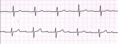
Difficulty transmitting impulse through AV node (where SA node is normal) – Due to this delay, the PR interval is longer – Once this travels through AV node, the impulse travels normally.
| QRS Complex | P Wave | PR Interval | Rate (bpm) |
|---|---|---|---|
| 0.08 – 0.10s (2 – 2.5 small boxes) | Present | >0.20s (> 5 small boxes) | 50 – 100 bpm |
- AMI
- Congenital abnormalities
- Degenerative changes
- Drug toxicity
- Ischaemic heart disease
- Vagal activity
- Nausea & Vomiting
- Related to ‘causes’ symptoms
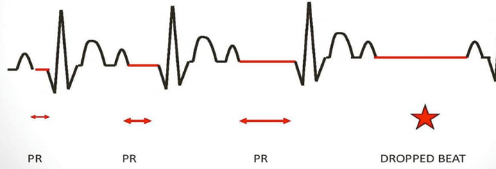
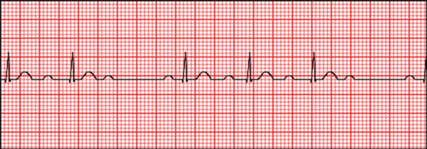
Represents a delayed transmission impulse through the AV node – However the time it takes to reach the ventricles increase for every impulse. This results in gradual PR interval increase. This continues until SA node intervenes with a sinus beats which resets the cycle again to prevent a fall in blood pressure.
- RR interval shortens with each beat of the cycle
- P-P interval remains constant
| QRS Complex | P Wave | PR Interval | Rate (bpm) |
|---|---|---|---|
| 0.08 – 0.10s (2 – 2.5 small boxes) | Present (more than QRS complex) | Progressive Prolongation | 50 – 100 bpm |
- AMI
- Cardiac infections
- Drug toxicity
- Ischaemic heart disease
- Increase vagal tone
- Lightheaded / dizzy
- Chest pain
- Regularly has irregular heartbeat
- Bradycardia
- Hypotension

Severe 2nd degree block resulting in a dropped beat – Due to SA node impulse not being transmitted to the ventricles, without progressive prolongation of the PR interval. If 2 or more impulses are blocked at the AV node before reaching the ventricles, it is called 2:1 or 3:2 block.
Can develop into 3rd degree AV block or ventricular standstill.
| QRS Complex | P Wave | PR Interval | Rate (bpm) |
|---|---|---|---|
| 0.08 – 0.10s (2 – 2.5 small boxes) Can sometimes widened | Present | Normal when P-wave & QRS are together | Can be slow |
- AMI
- Degenerative changes
- Drug toxicity
- Ischaemic heart disease
- Fall in blood pressure from dropped beat
- Syncope
- Chest pain
- Regularly has irregular heartbeat
- Bradycardia
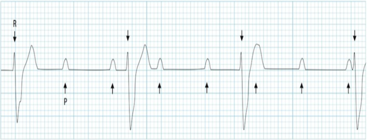
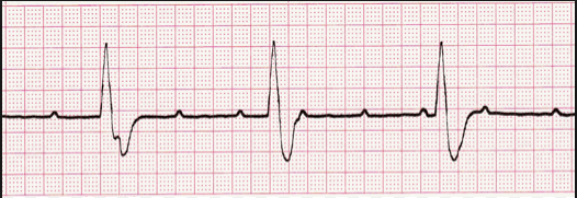

No communication through AV node resulting in separate atrial & ventricle transmission by their own branches. The SA node fires at 60-100 where the ventricles are at about 20-40bpm from nearby impulses from AV node or; within the ventricles.
Perfusion is through junctional or ventricular escape rhythm – Can result to ventricle standstill.
Hypotension is displayed due to inability of atria to intervene resulting in diminished cardiac output.
| QRS Complex | P Wave | PR Interval | Rate (bpm) |
|---|---|---|---|
| 0.08 – 0.10s (2 – 2.5 small boxes) Can sometimes widened | Normal but more than QRS complex | Dissociated relationship | Variable Atria (60-100bpm) and Ventricle (20-40bpm) rates. |
- AMI
- Degenerative changes
- Drug toxicity
- Ischaemic heart disease
- From other AV blocks
- Lightheaded / dizzy
- Syncope
- Chest pain
- Bradycardia
- •Hypotension
Page contributors:
 | Thanh Bui, AP60825 Event Medic, Emergency Medical Technician & Volunteer Development Officer
|
 | Andrew Moffat, AP16790 |
Clinical Resources Website
St John Ambulance Western Australia Ltd (ABN 55 028 468 715) (St John WA) operates ambulance and other pre-hospital clinical services. St John WA’s Clinical Resources, including its Clinical Practice Guidelines (Clinical Resources), are intended for use by credentialed St John WA staff and volunteers when providing clinical care to patients for or on behalf of St John WA, within the St John WA Clinical Governance Framework, and only to the extent of the clinician’s authority to practice.
Other users – Terms of Use
The content of the St John WA Clinical Resources is provided for information purposes only and is not intended to serve as health, medical or treatment advice. Any user of this website agrees to be bound by these Terms of Use in their use of the Clinical Resources.
St John WA does not represent or warrant (whether express, implied, statutory, or otherwise) that the content of the Clinical Resources is accurate, reliable, up-to-date, complete or that the information contained is suitable for your needs or for any particular purpose. You are responsible for assessing whether the information is accurate, reliable, up-to-date, authentic, relevant, or complete and where appropriate, seek independent professional advice.
St John WA expressly prohibits use of these Clinical Resources to guide clinical care of patients by organisations external to St John WA, except where these organisations have been directly engaged by St John WA to provide services. Any use of the Clinical Resources, with St John WA approval, must attribute St John WA as the creator of the Clinical Resources and include the copyright notice and (where reasonably practicable) provide a URL/hyperlink to the St John WA Clinical Resources website.
No permission or licence is granted to reproduce, make commercial use of, adapt, modify or create derivative works from these Clinical Resources. For permissions beyond the scope of these Terms of Use, including a commercial licence, please contact medservices@stjohnambulance.com.au
Where links are provided to resources on external websites, St John WA:
- Gives no assurances about the quality, accuracy or relevance of material on any linked site;
- Accepts no legal responsibility regarding the accuracy and reliability of external material; and
- Does not endorse any material, associated organisation, product or service on other sites.
Your use of any external website is governed by the terms of that website, including any authorisation, requirement or licence for use of the material on that website.
To the maximum extent permitted by law, St John WA excludes liability (including liability in negligence) for any direct, special, indirect, incidental, consequential, punitive, exemplary or other loss, cost, damage or expense arising out of, or in connection with, use or reliance on the Clinical Resources (including without limitation any interference with or damage to a user’s computer, device, software or data occurring in connection with such use).
Cookies
Please read this cookie policy carefully before using Clinical Resources from St John WA.
The cookies used on this site are small and completely anonymous pieces of information and are stored on your computer or mobile device. The data that the cookies contain identify your user preferences (such as your preferred text size, scope / skill level preference and Colour Assist mode, among other user settings) so that they can be recalled the next time that you visit a page within Clinical Resources. These cookies are necessary to offer you the best and most efficient possible experience when accessing and navigating through our website and using its features. These cookies do not collect or send analytical information back to St John WA.
Clinical Resources does integrate with Google Analytics and any cookies associated with this service enable us (and third-party services) to collect aggregated data for statistical purposes on how our visitors use this website. These cookies do not contain personal information such as names and email addresses and are used to help us improve your user experience of the website.
If you want to restrict or block the cookies that are set by our website, you can do so through your browser setting. Alternatively, you can visit www.internetcookies.com, which contains comprehensive information on how to do this on a wide variety of browsers and devices. You will find general information about cookies and details on how to delete cookies from your device. If you have any questions about this policy or our use of cookies, please contact us.