
Refers to abnormally of muscle cells resulting in large ventricles or atria resulting in reduced functions, creating potential arrythmias, caused by:
- Dilation by volume (diastolic) overload: Increased pressure within the chamber causing dilation; this is due to the regurgitation of blood due to impaired valves or inability to pump blood forward due to afterload (hypertension)
- Hypertrophy by pressure (systolic) overload: Enlargement of muscle cells due to increased demand or pressure which requires more muscle bulk & cell expansion (usually in athletes). Other reasons include increase resistance in the aortic valve making it difficult to pump blood towards. Other causes can include pulmonary disorders where pressure can backlog the heart.
- Increased QRS amplitudes: As muscle mass increase, so does electrical potentials therefore producing a stronger impulse, furthermore, larger chamber may be near the electrodes producing higher amplitude. Exception is late dilation where ventricular function is poor = lower QRS amplitude
- Prolongation of QRS duration: Slight change due to longer depolarisation time for larger mass – Should not exceed 0.12 seconds (unless due to intraventricular conduction defects).
- Axis changes: Can shift to right (RVH) or left (LVH)
- ST-T segment changes: Secondary ST-T changes such as ST elevation & depression can occur with presence of being discordant to the QRS complex in significant hypertrophy.
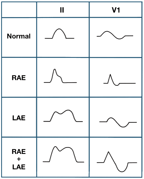
Can lead to having atrial fibrillation or flutter (AF) - Usually P-wave is impacted & examined in V1 + Lead II.
Usually from mitral valve impairment causing resistance to flow, hence enlarging the left atrium – A second hump in P-wave is seen showing right atrium contraction before the left.
ECG Findings
- Biphasic p-wave in V1 with wide, deep (> 1mm) terminal component
- Left atrial hyper-terminal component is larger than .04 sec
Pressure in the pulmonary system or resistance in valve can cause right atrium hypertrophy in order to pump blood to the ventricles. This generates a stronger impulse (hence higher P-wave amplitude) where the wave can be seen as a sharp peak – P-wave amplitude is > 2.5mm
ECG Findings
- Rarely isolated finding (usually RVH/RAD also)
- P amplitude >2.5mm in II
- Large biphasic p wave in V1 (> 1.5 mm in V1 and V2)
- Right atrial hyper.-initial component is larger in V1 than V6
- p-mitrale-m notched p wave in leads 1 and 2. Greater than .12 seconds.
Implies enlargement in both left & right atrium (seen as large P-wave in Lead II, and large biphasic P-wave in V1).
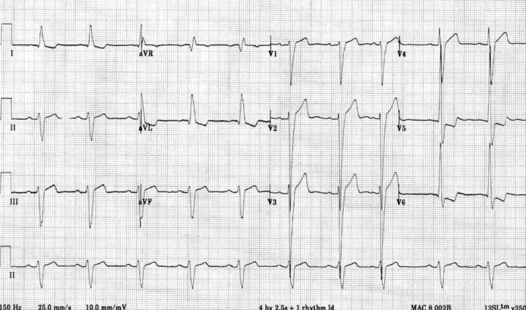
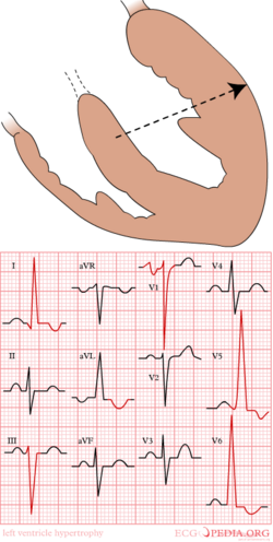
Also known as left/right ventricular hypertrophy (LVH/RVH), can lead to having ventricular tachycardia (VT) – Usually QRS is impacted (often tall QRS complex due to increased muscle mass).
Usually V1-V2, & V5-V6 are impacted – Generally by aortic stenosis, insufficiency or hypertension, overloading causes broader QRS where longer depolarisation occurs in the left ventricles.
- R-Wave: V5-V6, Lead I, & aVL show larger R-waves – Usually > 30mm in V5
- S-Wave: Deeper S-waves shown in V1-V2 due to amplified vector of left ventricles
- QRS Complex: Can be notched and prolonged in duration
- V5-V6 Secondary ST-T: ST downslope & T-wave inversion can occur
- V5-V6 Secondary ST-T: ST elevation can occur, ST segment is concaved
- P-mitral may be present (as it affects hemodynamic of left atrium
- Left axis deviation may be present
- Longer QTc interval
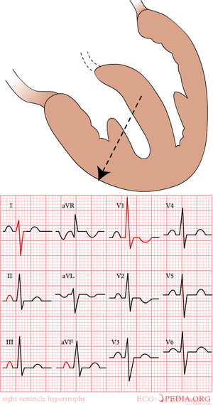
Usually, left ventricle’s vector is larger than the right, therefore QRS is dominated by left ventricle. However, right ventricle can be seen in QES due to hypertrophy – This can be by lung or heart disease, and valve impairment.
- R-Wave: V1 & V2 show larger R-waves, wave progression is smaller from V1 to V6
- S-Wave: Small S-wave observed in V1
- QRS Complex: Prolonged slightly (< 120ms) where R-wave is 35-55ms in V1-V2
- V1-V3 Secondary ST-T: ST-T is discordant to QRS complex
- rSR’ pattern can be seen in V1 & V2, can resemble RBBB, but is not
- P-pulmonale can be present
- Electrical axis almost resembles RAD
- V5-V6, Lead I, & aVL display smaller R-waves; due to opposite R-wave progression from V1 to V6
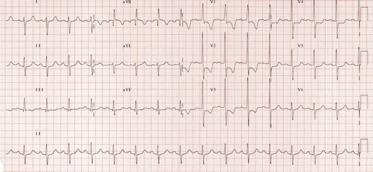
Detection is low to detect biventricular hypertrophy due to both vectors cancelling each other out when both vectors are hypertrophied.
When LVH is shown, an examination should occur to see if RVH is present - shown by:
- Right axis deviation – This never occurs in LVH
- Deep S-wave (> 6mm) in V5 or V6
- Large RS-complex in multiple leads
- P-pulmonale present
Page contributors:
 | Thanh Bui, AP60825 Event Medic, Emergency Medical Technician & Volunteer Development Officer
|
 | Andrew Moffat, AP16790 |
Clinical Resources Website
St John Ambulance Western Australia Ltd (ABN 55 028 468 715) (St John WA) operates ambulance and other pre-hospital clinical services. St John WA’s Clinical Resources, including its Clinical Practice Guidelines (Clinical Resources), are intended for use by credentialed St John WA staff and volunteers when providing clinical care to patients for or on behalf of St John WA, within the St John WA Clinical Governance Framework, and only to the extent of the clinician’s authority to practice.
Other users – Terms of Use
The content of the St John WA Clinical Resources is provided for information purposes only and is not intended to serve as health, medical or treatment advice. Any user of this website agrees to be bound by these Terms of Use in their use of the Clinical Resources.
St John WA does not represent or warrant (whether express, implied, statutory, or otherwise) that the content of the Clinical Resources is accurate, reliable, up-to-date, complete or that the information contained is suitable for your needs or for any particular purpose. You are responsible for assessing whether the information is accurate, reliable, up-to-date, authentic, relevant, or complete and where appropriate, seek independent professional advice.
St John WA expressly prohibits use of these Clinical Resources to guide clinical care of patients by organisations external to St John WA, except where these organisations have been directly engaged by St John WA to provide services. Any use of the Clinical Resources, with St John WA approval, must attribute St John WA as the creator of the Clinical Resources and include the copyright notice and (where reasonably practicable) provide a URL/hyperlink to the St John WA Clinical Resources website.
No permission or licence is granted to reproduce, make commercial use of, adapt, modify or create derivative works from these Clinical Resources. For permissions beyond the scope of these Terms of Use, including a commercial licence, please contact medservices@stjohnambulance.com.au
Where links are provided to resources on external websites, St John WA:
- Gives no assurances about the quality, accuracy or relevance of material on any linked site;
- Accepts no legal responsibility regarding the accuracy and reliability of external material; and
- Does not endorse any material, associated organisation, product or service on other sites.
Your use of any external website is governed by the terms of that website, including any authorisation, requirement or licence for use of the material on that website.
To the maximum extent permitted by law, St John WA excludes liability (including liability in negligence) for any direct, special, indirect, incidental, consequential, punitive, exemplary or other loss, cost, damage or expense arising out of, or in connection with, use or reliance on the Clinical Resources (including without limitation any interference with or damage to a user’s computer, device, software or data occurring in connection with such use).
Cookies
Please read this cookie policy carefully before using Clinical Resources from St John WA.
The cookies used on this site are small and completely anonymous pieces of information and are stored on your computer or mobile device. The data that the cookies contain identify your user preferences (such as your preferred text size, scope / skill level preference and Colour Assist mode, among other user settings) so that they can be recalled the next time that you visit a page within Clinical Resources. These cookies are necessary to offer you the best and most efficient possible experience when accessing and navigating through our website and using its features. These cookies do not collect or send analytical information back to St John WA.
Clinical Resources does integrate with Google Analytics and any cookies associated with this service enable us (and third-party services) to collect aggregated data for statistical purposes on how our visitors use this website. These cookies do not contain personal information such as names and email addresses and are used to help us improve your user experience of the website.
If you want to restrict or block the cookies that are set by our website, you can do so through your browser setting. Alternatively, you can visit www.internetcookies.com, which contains comprehensive information on how to do this on a wide variety of browsers and devices. You will find general information about cookies and details on how to delete cookies from your device. If you have any questions about this policy or our use of cookies, please contact us.